Unmixing Fluorescent Channels for Breast Cancer Metabolic Imaging
Roarke Horstmeyer Sarah Mekha Katie Broun Marcia dos Santos
rwh4@duke.edu sarah.mekha@duke.edu katie.broun@duke.edu mc884@duke.edu
| Paper PDF |
|
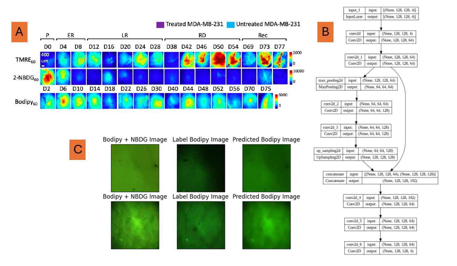
Breast cancer is the most common cancer in women worldwide; the most aggressive form of triple negative breast cancer lacks many hormone receptors making tracking this overall disease and tumor molecular subpopulations. Our lab has developed an optical toolkit of three fluorophores to measure metabolic changes over time. Given the optical crosstalk of some of our fluorophores, our goal was to be able to image metabolic features simultaneously in vivo, therefore needing unmixing capabilities, which we hope to achieve with a convolutional neural network model. We used different combinations of our simulated RGB images to perform several experiments of unmixing two or three channels. We found that more distinct channels (green and red) gave better results than overlapping channels (green and green). Landmark features were visually observed but at a lower resolution and contrast than the true labels. We believe more reliable results can be achieved with more structural and optical information. The figure shows (A) the raw data collected by Sunassee ED, (B) the modified U-Net architecture used in the project, and (C) the results of the network trained on Bodipy and 2-NBDG mixed into an RGB image and with Bodipy as a label.
Paper:
|
Reference papaer
|
Data
| |