MRI Image Healthy Abdominal Organ Segmentation with a U-Net model
Jing LI Yirui XU
jl1104@duke.edu yx219@duke.edu
| Paper PDF |
|
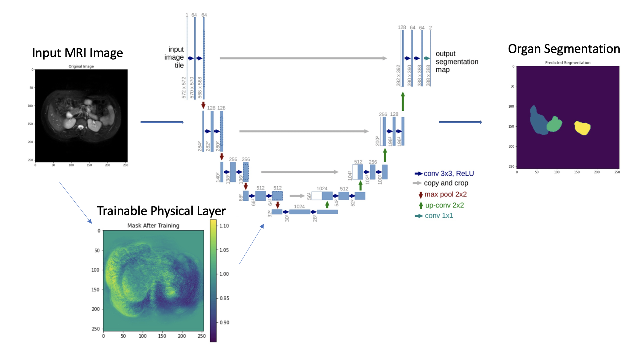
Medical image segmentation is the key issue to determine whether medical image can provide reliable basis in clinical diagnosis and treatment. In recent years, medical image segmentation technology has made remarkable progress due to the application of deep learning algorithm in medical image segmentation. After the U-Net network was proposed, it performed well in the field of medical image segmentation. In this project, we presented a U-Net implementation with a trainable mask physical layer for the segmentation of healthy abdominal organs in MRI images. For liver segmentation, the validation accuracy was 72.4% without the physical and was 74.5% with the physical layer, indicating that the trainable physical layer improved the model performance. In both cases, there was a gap between training accuracy and validation accuracy. We also implemented single organ segmentation to the right kidney, left kidney, and spleen, and combined the single-organ mask for the segmentation of healthy abdominal organs. The dice coefficient of predicted segmentation is 82.2%. To improve the performance of the model, future work such as increasing data size, and using dice coefficient as loss function should be done.
|
| Paper: |
| Code and Data: |